International Journal of Cardiovascular Sciences. 19/fev/2025;38:e20230170.
Electrocardiographic Analysis of Ventricular Repolarization in Patients With Psoriasis: A Cross-Sectional Study
Guilia Bevilacqua Schmitz
![]() , Raíssa Massaia Londero Chemello
, Raíssa Massaia Londero Chemello
![]() , Marco Lumertz Saffi
, Marco Lumertz Saffi
![]() , Luciane Prado de Vargas
, Luciane Prado de Vargas
![]() , Diego Chemello
, Diego Chemello
![]()
Abstract
Background
Psoriasis is a chronic inflammatory disease. Cardiovascular diseases, including arrhythmias, tend to occur more frequently in these patients.
Objective
This study aimed to investigate new electrocardiographic markers for assessing ventricular repolarization in outpatients with psoriasis in a tertiary center in Brazil.
Methods
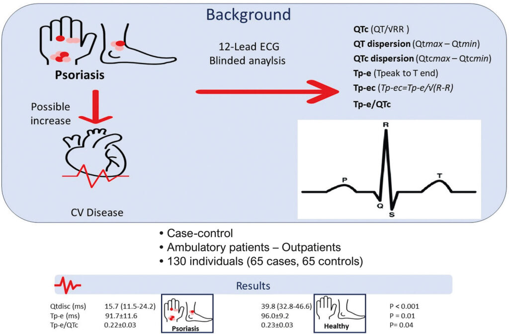
This work is a cross-sectional study, which selected outpatients with psoriasis undergoing follow-up in a tertiary dermatology service in southern Brazil. The controls were selected from a private outpatient clinic. All the electrocardiogram (ECG) analyses were performed by one certified electrophysiologist. Comparative statistical tests and Spearman’s correlation analysis were used. Measures with statistically significant differences in the univariate analysis were taken to the multivariate analysis (Generalized Linear Model – Poisson Regression with age adjustment). A P<0.05 value was considered statistically significant for all analyses.
Results
A total of 130 individuals were evaluated, mean age of 53.1±15.1 years. The univariate analysis showed no difference in Corrected QT interval (QTc) between the groups, considering 411.2±21.2 ms in the psoriasis group (PG) versus 412.8±25.2 ms in the control group (CG) (p = 0.694). The median values of Corrected QT dispersion (QTdisc) were lower in the PG as compared to the CG, 15.7 ms (IQR 11.5–24.2 ms) versus 39.8 ms (Interquartile Range, IQR, 32.8–46.6 ms), respectively (p<0.001). For peak-to-end interval of the T wave (Tp-e) and Tp-e/QTc values, the mean values were also lower in patients with psoriasis. For Tp-e, the mean values in the PG were 91.7±11.6 ms versus 96.0±9.2 ms in the CG (p = 0.024). For The Tp-e/QTc, the mean values in the PG were 0.22±0.03 versus 0.23±0.03 in the CG (p = 0.024). The multivariate analysis, adjusted for age, QTdis, Qtdisc, Tp-e, and Tp-e/QTc, remained independently lower in patients with psoriasis.
Conclusion
The evaluation of ventricular repolarization parameters in a sample of outpatients with psoriasis in southern Brazil showed that ventricular repolarization values were generally lower when compared to controls. No significant correlation was found between disease activity and ventricular repolarization parameters.
Palavras-chave: psoriasis; electrocardiography; cardiac arrhythmias; ventricular tachycardia
277