International Journal of Cardiovascular Sciences. 04/maio/2022;35(5):676-80.
Pacemaker Implantation without Fluoroscopy and Guided by Anatomical Mapping
Mauricio Montemezzo
![]() , Ahmed AlTurki
, Ahmed AlTurki
![]() , Marcos Jakolinski
, Marcos Jakolinski
![]() , Jose Carlos Moura Jorge
, Jose Carlos Moura Jorge
![]()
Introduction
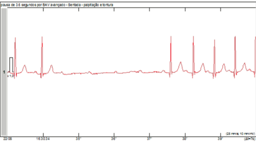
Cardiogenic syncope is an uncommon pathology in the context of pregnancy, and an associated transient atrioventricular block is even more rare. The harmful effects of radiation exposure for fetus are already well-known. Thus, alternative guiding techniques are being proposed as an alternative in order to avoid radiation exposure. Previous reports have described the use of intracardiac echocardiography or three-dimensional (3D) electroanatomical mapping. Despite efforts to develop non-fluoroscopic approaches, cardiac device implantation still requires some fluoroscopic imaging. Each hour of fluoroscopic imaging is estimated to increase the lifetime risk of developing a fatal malignancy by up to 1%, as well as being an increased risk of a genetic defect in up to 20 in every 1 million births. ,
With technological advancement, the use of less invasive techniques, together with short procedure times and less radiation exposure is essential. Therefore, repurposing technologies, which were originally developed for other applications, but that have been proven to be safe, should be encouraged. In this light, the present study advocates the use of the CARTO system technology (Biosense Webster, INC., Diamond Bar, CA) to delineate right atrium and right ventricle geometry so as to allow the pacemaker lead to be identified as a catheter to navigate the cardiac cavity without fluoroscopic exposure.
[…]
Palavras-chave: Artificial Pacemaker; Fluoroscopy; Pregnancy
887